Hysteroscopy: What It Is, What It Finds, and When You Need It
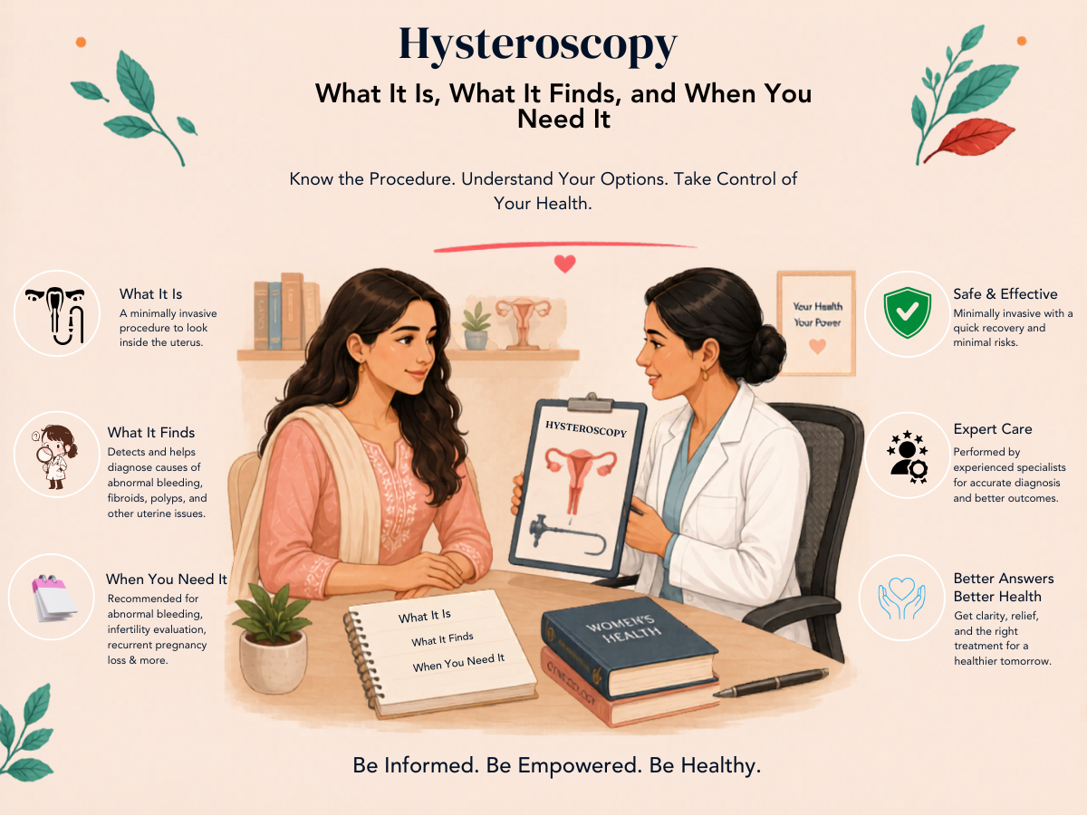
Hysteroscopy — direct camera examination of the uterine cavity — is both a diagnostic tool and a therapeutic procedure. It is the gold standard investigation for conditions affecting the inside of the uterus, and it allows simultaneous treatment of many conditions it finds — polyp removal, fibroid resection, septum division, adhesion release — in a single minimally invasive procedure. Understanding what hysteroscopy involves, when it is indicated, and what to expect helps women approach it with confidence rather than anxiety.
What Is Hysteroscopy?
A hysteroscope is a thin, rigid or flexible optical instrument (typically 3 to 5 mm in diameter) that is passed through the cervical canal into the uterine cavity. The cavity is distended with fluid (normal saline or glycine) or gas (CO2 for office procedures) to open it for clear visualisation. The interior of the uterine cavity — including the endometrial lining, the tubal openings (ostia), and any structural abnormalities — is directly seen on a monitor.
Hysteroscopy is performed in two settings:
- Office/outpatient hysteroscopy: Using a very fine hysteroscope (2 to 3 mm), typically without anaesthesia or with local anaesthetic. Suitable for diagnostic assessment and very small procedures (minor polyp sampling, IUS insertion under vision).
- Operative hysteroscopy: Under general or regional anaesthesia, using a resectoscope (an instrument with a cutting element) or other operative devices. Allows removal of polyps, submucosal fibroids, uterine septa, and adhesions.
What Hysteroscopy Can Find
- Endometrial polyps: Localised overgrowths of the endometrial lining — often missed on standard ultrasound, clearly visible at hysteroscopy.
- Submucosal fibroids: Fibroids protruding into the uterine cavity — hysteroscopy characterises their size and degree of cavity involvement (classification relevant to resectability).
- Uterine septum: A congenital fibrous band dividing the cavity — the most common correctable structural cause of recurrent pregnancy loss. Hysteroscopic metroplasty (septum division) is a day procedure with excellent outcomes.
- Intrauterine adhesions (Asherman's syndrome): Fibrous bands or sheets scarring the cavity after previous uterine surgery or infection. Hysteroscopic adhesiolysis releases these.
- Endometrial abnormalities: Irregular or thickened areas of endometrium that may indicate hyperplasia or malignancy — targeted biopsy can be taken under direct vision.
- Tubal ostia: Whether both tubal openings are visible, which can provide indirect evidence about tubal anatomy.
- Congenital uterine anomalies: Bicornuate or unicornuate uterus may be identified.
When Is Hysteroscopy Indicated?
Before IVF
Hysteroscopy before IVF is a contentious but practically important question. Routine hysteroscopy before every first IVF cycle — in the absence of any specific indication — has not been shown to improve IVF success rates in all populations (the Investigate clinical trial in the UK showed no benefit in unselected patients).
However, hysteroscopy is appropriate before IVF in:
- Unexplained IVF failure — after two or more failed cycles despite good embryo quality
- Abnormal findings on uterine assessment (ultrasound, HyCoSy, or SIS) suggesting polyp, fibroid, or cavity distortion
- History of recurrent pregnancy loss — to detect and treat septum, adhesions, or other cavity abnormalities
- Suspected Asherman's syndrome or thin endometrium not responding to standard preparation
- Any clinical indication for cavity evaluation regardless of IVF status
For Heavy Periods or Abnormal Bleeding
Hysteroscopy is the gold standard investigation for abnormal uterine bleeding — providing the direct visualisation that accurately characterises the cause and allows simultaneous treatment (polypectomy, myomectomy) if a lesion is found.
For Recurrent Pregnancy Loss
All women with two or more consecutive miscarriages should have their uterine cavity assessed — ideally by hysteroscopy or saline sonography. A uterine septum, intrauterine adhesions, or significant cavity distortion from fibroids are correctable causes of recurrent loss.
What to Expect from the Procedure
Under anaesthesia (for operative hysteroscopy):
- Fasting from midnight before the procedure
- Procedure takes 15 to 45 minutes depending on whether it is diagnostic or operative
- Recovery in the day procedure area for 1 to 3 hours
- Mild cramping and light spotting for 1 to 2 days after the procedure — normal and expected
- Return to normal activities within 24 to 48 hours for diagnostic procedures; 1 week for operative procedures
- Abstain from intercourse and tampons for 2 to 4 weeks after operative procedures to allow the cavity to heal
Hysteroscopy and Fertility
When a hysteroscopy finds and treats a uterine lesion — a polyp, a submucosal fibroid, a septum, adhesions — the impact on fertility can be significant:
- Polypectomy: Small, well-designed studies show improved pregnancy rates after removal of endometrial polyps in infertile women.
- Submucosal fibroid resection: Significantly improves implantation rates in IVF — some studies showing restoration to rates equivalent to fibroid-free women.
- Septum resection: Reduces miscarriage rates significantly in women with recurrent pregnancy loss and a uterine septum.
- Adhesiolysis for Asherman's syndrome: Restores fertility in mild to moderate cases; severe Asherman's syndrome is much harder to treat and may require repeated procedures.
Frequently Asked Questions
Q1. Is hysteroscopy painful?
Operative hysteroscopy under general anaesthesia is not experienced during the procedure. Office or outpatient hysteroscopy (without anaesthesia) is performed with a very fine instrument and is tolerated by most women with mild to moderate cramping — comparable to period pain. Pre-medication with ibuprofen 30 to 60 minutes before the procedure significantly reduces discomfort. Anxious patients or those with cervical stenosis may be offered sedation or general anaesthesia even for diagnostic procedures.
Q2. Can a hysteroscopy fix thin endometrium?
Hysteroscopy can identify causes of thin endometrium — such as intrauterine adhesions that reduce the functional endometrial surface — and treat them. Adhesiolysis for Asherman's syndrome is one of the most important treatments for thin endometrium in affected women. However, if the thin endometrium is from radiation damage, severe Asherman's, or intrinsic endometrial insufficiency, hysteroscopy alone may not restore normal lining — additional interventions (high-dose oestrogen, PRP infusion) may be needed alongside.
Q3. I had a hysteroscopy 6 months ago and a polyp was removed. Do I need another one before IVF?
If the polyp was fully removed and confirmed on the operative report, a repeat hysteroscopy within 6 months is not routinely needed before IVF. If you have had ongoing symptoms (irregular bleeding, heavy periods) since the procedure, or if the ultrasound before your IVF cycle shows any concern, a reassessment would be appropriate. In the absence of new symptoms or findings, proceeding with IVF on the basis of the previous successful polypectomy is reasonable.
DISCLAIMER: This article is for educational purposes only and does not constitute medical advice. Consult Dr. Sunita Tandulwadkar or a qualified specialist for personalised guidance. Solo Clinic IVF & ObGyn, Pune.